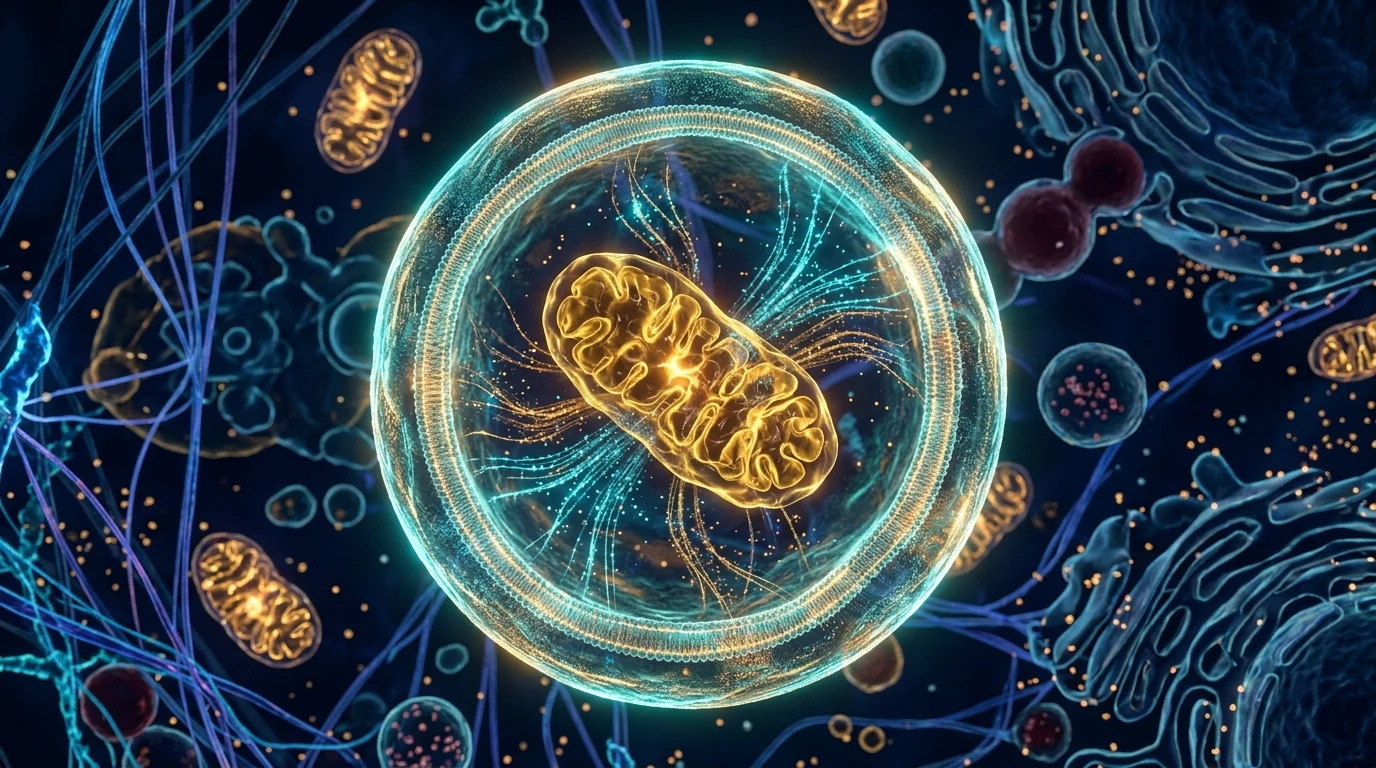
- → 2016 Nobel Discovery: Yoshinori Ohsumi elucidated the core ATG gene family that orchestrates how cells parcel and recycle their own molecular debris.
- → Autophagosome-Lysosome Fusion: The phagophore membrane sequesters damaged organelles within a double-membraned autophagosome; acidic lysosomal hydrolases then digest this cargo into amino acids and fatty acids.
- → Mitophagy and Energy Restoration: Dysfunctional, ROS-leaking mitochondria are selectively eliminated, preserving cellular vitality and metabolic fitness.
- → Spermidine and Fasting Synergy: Intermittent fasting and dietary polyamines like spermidine stimulate autophagic flux, promoting healthy cellular longevity.
📌 Cellular Rejuvenation and Autophagy Series — Part 3/3 (Final)
- Part 1: The Autophagy Clock: When Does Cellular Recycling Actually Peak During Intermittent Fasting?
- Part 2: 5 Hidden Mistakes That Halt Autophagy: The Collagen, Milk Coffee, and Sweetener Trap
- Part 3 (You are here): Nobel-Prize Winning Waste Disposal: The Biochemistry of Cellular Recycling
One of the foundational drivers of biological aging is not simply cellular chronological wear, but the intracellular accumulation of un-cleared molecular debris.
Over decades, misfolded protein aggregates, exhausted organelles, and damaged, free-radical-leaking mitochondria clutter the cytoplasm. Japanese cell biologist Yoshinori Ohsumi elucidated the genetic machinery behind how cells systematically package, degrade, and recycle this cellular waste into metabolic fuel, earning the 2016 Nobel Prize in Physiology or Medicine [1].
In this final installment of our series, we dissect the molecular gears of the autophagic recycling machinery and examine its evidence-based impact on longevity.
1. Visualizing the Process: The Cellular Compactor and Recycling Conveyor
To grasp how intracellular sanitation operates, visualize a three-stage biochemical recycling line:
[Stage 1: Phagophore] ── Crescent-shaped isolation membrane nucleates
        │
        ▼
[Stage 2: Autophagosome] ── Double-membrane vesicle engulfs toxic debris and organelles
        │
        ▼
[Stage 3: Autophagolysosome] ── Acidic lysosomal hydrolases digest cargo into building blocks- Step 1 — Sequestration (The Phagophore): When intracellular energy sensors register nutrient depletion (AMPK activation and mTORC1 suppression), a de novo, crescent-shaped isolation membrane (the phagophore) nucleates in the cytoplasm [1].
- Step 2 — Packaging (The Autophagosome): This membrane elongates, engulfing damaged organelles, misfolded protein clusters, and worn-out structures, sealing itself into a spherical double-membrane vesicle called the autophagosome [1].
- Step 3 — Digestion and Reclaiming (The Autophagolysosome): The autophagosome docks and fuses with a lysosome, the cell’s degradation center. Acidic hydrolases break down the captured cargo into fundamental metabolic building blocks: free amino acids, fatty acids, and nucleotides [1].
The cell immediately repurposes these raw recycled substrates to synthesize fresh functional proteins and refuel mitochondrial oxidative phosphorylation [1], [2].
2. Yoshinori Ohsumi’s Breakthrough: Why the ATG Genes Matter
Ohsumi’s pioneering work in yeast demonstrated that cellular self-digestion is not a disordered death pathway, but a tightly orchestrated, genetically conserved cascade [1].
He identified and characterized the core ATG (Autophagy-Related) gene family, which governs every sequential step of autophagic flux—from phagophore initiation and membrane elongation to vesicle closure and lysosomal fusion [1]. Through this molecular mechanism, the cell selectively isolates and cleans cytotoxic intracellular aggregates [1].
Constitutive basal autophagy operates continuously to prevent proteotoxicity, even in the fed state [2]. When autophagic flux is impaired, cytotoxic protein aggregates accumulate, accelerating cellular senescence and tissue degeneration [2]. Conversely, robust autophagic capacity bolsters cellular stress resistance, genomic integrity, and healthy longevity [2].
3. Mitophagy: Mitochondrial Quality Control
Mitochondria generate adenosine triphosphate (ATP) through oxidative phosphorylation. However, this electron transport process generates an inevitable byproduct: Reactive Oxygen Species (ROS / Free Radicals) [3].
With persistent oxidative exposure, mitochondrial inner membranes lose electrical potential ($\Delta\Psi_m$), rendering them inefficient and excessively leakier of ROS [3]. This is where mitophagy—the selective autophagic degradation of mitochondria—becomes indispensable:
- Targeted Tagging: The cell identifies depolarized, defective mitochondria via the PINK1-Parkin signaling pathway, ubiquitously marking their outer membranes [3].
- Selective Sequestration: Autophagosomal receptors recognize these ubiquitin tags, specifically enveloping the dysfunctional organelle while sparing healthy mitochondria [3].
- Cellular Revitalization: Degrading defective units reduces systemic oxidative stress and preserves overall cellular metabolic efficiency [3], [5]. For a detailed scientific evaluation of commercial supplements and controversial longevity remedies aimed at mitochondrial optimization, see our comprehensive guide on Molecular Hydrogen, Methylene Blue, and Heavy Metal Detox Realities.
A landmark review in Nature Cell Biology (Palikaras et al., 2018) underscored that efficient mitophagy is an absolute requirement for sustaining neuronal viability, cardiac function, and metabolic homeostasis throughout aging [3].
4. Spermidine and Longevity: The Endogenous Autophagy Inducer
Nutrient deprivation is not the sole physiological driver of autophagic cleansing. Molecular research has identified endogenous compounds that directly stimulate this cascade:
- Endogenous Stimulator (Spermidine): In a seminal study published in Nature Cell Biology (Eisenberg, Madeo et al., 2009), the natural polyamine spermidine was demonstrated to potently induce autophagy, reduce reactive oxygen species, and extend healthy lifespan in multiple model organisms [4].
- Fasting-Spermidine Synergy: Periods of caloric restriction stimulate endogenous polyamine synthesis, producing a synergistic amplification of cellular self-clearing mechanisms [4], [5].
5. Clinical and Integrative Medicine Perspectives: Restoring Biological Clearance
Integrative physicians and clinical researchers (such as Dr. med. Petra Wiechel and René Gräber, featured on Swiss health channel QS24) frame autophagy as humanity’s most primal evolutionary shield against chronic metabolic overload [6]:
- Creating Biological Clearance Windows: Continuously bombarding cells with nutrients keeps anabolic pathways locked on. Providing intermittent digestive pauses creates the metabolic space required for cellular debris clearance [6].
- Hydration and Metabolic Flux: Consuming pure mineral water and unsweetened herbal infusions during fasting windows facilitates the renal elimination of metabolized waste without triggering nutrient sensors [6].
6. Series Action Plan: The Cellular Rejuvenation Protocol
To translate the scientific insights across our three-part series into daily practice:
- Target the 16-Hour Fasting Threshold: Autophagic flux accelerates significantly between the 14th and 16th fasting hours (Part 1).
- Safeguard the Fasting Window: Reserve collagen peptides, BCAAs, milk, and synthetic sweeteners strictly for your eating window (Part 2).
- Stimulate Mitophagy: Pair intermittent fasting intervals with light to moderate physical activity to facilitate the turnover of damaged mitochondria (Part 3).
- Break the Fast Mindfully: Initiate your refeeding with steamed fiber-rich vegetables and healthy lipids to avoid acute postprandial glucose surges (QS24 Clinical Rule).
⚖️ Safety & Clinical Guidance:
Intermittent fasting and autophagic stimulation protocols are lifestyle optimization strategies intended for healthy adults. Individuals with chronic medical conditions, metabolic disorders, pregnant or lactating women, or those taking prescribed medications should never embark on extended fasting protocols without direct medical supervision.
Scientific Methodology and Primary Citations
- Ohsumi, Y. (2014). Historical landmarks of autophagy research. Cell Research, 24(1), 9–23. DOI: 10.1038/cr.2013.169 | PMID: 24366340
- Mizushima, N., & Komatsu, M. (2011). Autophagy: renovation of cells and tissues. Cell, 147(4), 728–741. DOI: 10.1016/j.cell.2011.10.026 | PMID: 22078877
- Palikaras, K., Lionaki, E., & Tavernarakis, N. (2018). Mechanisms of mitophagy in cellular homeostasis, physiology and pathology. Nature Cell Biology, 20(9), 1013–1022. DOI: 10.1038/s41556-018-0176-2 | PMID: 30154567
- Eisenberg, T., Knauer, H., Schauer, A., Madeo, F., et al. (2009). Induction of autophagy by spermidine promotes longevity. Nature Cell Biology, 11(11), 1305–1314. DOI: 10.1038/ncb1975 | PMID: 19801973
- de Cabo, R., & Mattson, M. P. (2019). Effects of Intermittent Fasting on Health, Aging, and Disease. New England Journal of Medicine, 381(26), 2541–2551. DOI: 10.1056/NEJMra1905136 | PMID: 31881139
- QS24 Schweizer Gesundheitsfernsehen. (2023–2024). Zellregeneration, Autophagie und Langlebigkeit (Dr. med. Petra Wiechel, René Gräber). QS24 TV Archiv & Mediathek.
For informational purposes only; consult a physician for medical diagnosis or treatment.