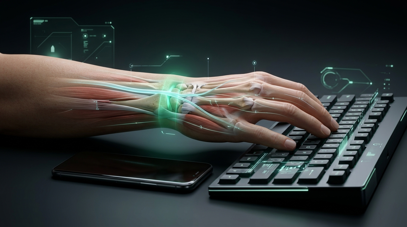
- → Desk and Keyboard Pressure: Pressing wrists against a hard desk during typing elevates intracarpal canal pressure well above the 30 mmHg microvascular ischemia threshold.
- → Smartphone Thumb Strain: Reaching for upper screen corners one-handed with large phones creates repetitive shear strain on thumb tendons, predisposing to De Quervain tenosynovitis.
- → Vertical Mouse Advantage: Maintaining the forearm in a neutral 57° handshake angle eliminates forearm pronation crossing (radius over ulna), dropping wrist torque and nerve compression.
- → Night Blue Light and Tendon Repair: Late-night screen exposure suppresses circadian melatonin secretion, blunting nighttime tenocyte cellular repair and collagen cross-linking.
⚠️ Important Clinical Note: This guide is strictly for informational and educational purposes. If you experience nocturnal wrist pain that awakens you from sleep, progressive hand numbness, grip weakness, or persistent paresthesia, consult an orthopedic hand surgeon, neurologist, or physical rehabilitation physician.
What Does This Guide Cover?
If your daily life involves typing on keyboards, clicking a mouse, gripping large smartphones, or operating handheld tools, your hands and wrists continuously endure cumulative mechanical microtrauma. Most individuals fail to notice this strain—until a persistent pinch flares at the base of the thumb, fingertips tingle, or nocturnal numbness disrupts deep sleep.
Modern life presents two predominant repetitive strain patterns:
- Smartphone Thumb & Pinky Resting Strain: One-handed handling of 200–240 gram smartphones forces the thumb to repeatedly reach across wide touchscreens while the pinky finger bears the device’s base weight (“Smartphone Pinky”).
- Desk, Keyboard & Mouse Ergonomics: Pressing carpal bones against hard desk edges, sustained forearm pronation (turning palms flat downward), and executing thousands of repetitive micro-clicks every shift.
Below, we examine the functional anatomy of the human wrist, the biomechanical pathophysiology of Carpal Tunnel Syndrome (CTS) and De Quervain tenosynovitis, and actionable, evidence-based ergonomic and biological prevention strategies.
💡 Authentic Biohacking: The most powerful biohack is never an artificial shortcut; it is the deliberate adoption of micro-habits that reduce physical joint torque, optimize posture, and accelerate intrinsic cellular tissue repair.
1. Biomechanics of the Wrist: How Mechanical Torque Generates Strain
The human wrist joint complex (articulatio radiocarpalis and mediocarpalis) represents an intricate kinematic chain designed to position the hand across three-dimensional space. The external static torque ($T$) exerted onto the wrist by a handheld smartphone, mouse, or handheld barcode scanner is governed by classical mechanics:
Static Torque Equation:
$T = F \times r \times \sin(\theta)$
Where:
- $T$ (Torque): Net rotational moment generated around the wrist joint (Newton·meters [N·m]),
- $r$ (Moment Arm): Perpendicular distance from the anatomical center of rotation (the capitate bone pivot) to the device’s Center of Mass ($CoM$) in meters [4],
- $F$ (Force): Gravitational payload of the device or keystroke/trigger actuation force ($F = m \cdot g$),
- $\theta$ (Angle): Angle between the force vector and the moment arm vector.
       Moment Arm (r: 70 - 140 mm)
◄─────────────────────────────────────────────►
[Wrist Pivot]                            [Center of Mass]
(Os Capitatum)                           (Phone / Mouse / Tool)
      O══════════════════════════════════════════■
      ▲                                          │
      │                                          │ F = m · g (Gravitational Force)
      │                                          ▼
Resistance Torque (T)
1.1. Daily Ergonomic Scenarios: Smartphones, Keyboards, and Scanners
To understand how cumulative mechanical stress transitions into chronic microvascular and tendon damage, let us evaluate three typical modern scenarios:
- Scenario A (Large Smartphone): Holding a 220 g smartphone (~2.16 N weight) with one hand places its center of mass approximately 10 cm (0.10 m) distal to your wrist pivot. The static torque countering gravity is: $T = 2.16\text{ N} \times 0.10\text{ m} = 0.22\text{ N}\cdot\text{m}$. While this figure appears modest, when the thumb hyper-abducts to reach the opposite upper screen corner, this lever arm exerts high shear stress across the basal first carpometacarpal (CMC) joint and extensor tendons.
- Scenario B (Office Keyboard & Mouse): An office worker logs an average of 15,000 to 30,000 keystrokes and 3,000 to 5,000 mouse clicks per 8-hour shift. Although key actuation requires only 1.5 to 3 N of force, performing these cycles with wrists compressed against a desk generates friction, micro-edema, and elevated hydrostatic pressure within the carpal canal [3, 9].
- Scenario C (Industrial Handheld Scanners & Barcode Terminals): Logistics warehouse workers operating 500–700 g scanners and pulling triggers 3,000 to 6,000 times daily generate instantaneous torques exceeding 1.05 N·m, pushing cumulative moment impulses well beyond occupational safety thresholds [3, 9].
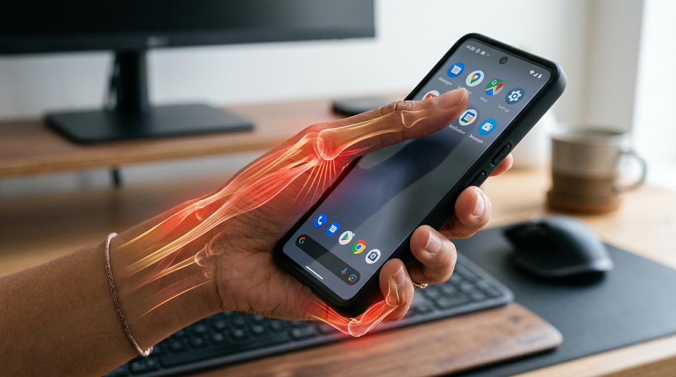 Figure 1: Biomechanical torque and tendon shear stress generated during single-handed smartphone use, showing extreme thumb excursion and pinky base shelf support (“Smartphone Pinky”).
📌 Fundamental Biomechanical Principle: Tissue micro-trauma depends not only on load magnitude, but critically on repetition frequency, joint deviation angles, and time spent under load (Cumulative Moment Impulse: $J_\tau = \int |T(t)|,dt$).
2. Intracarpal Hydrodynamic Pressure and Median Nerve Ischemia
2.1. Kinematic Coupled Motion of the Wrist
Radial deviation (angling toward the thumb) and ulnar deviation (angling toward the pinky) do not operate independently of flexion (bending forward) and extension (bending back).
In three-dimensional kinematic cadaveric and in vivo evaluations by Li et al. (2005), the anatomical geometry of the carpal rows and ligamentous constraints dictates mandatory coupled motion [3]:
- Extension + Radial Deviation
- Flexion + Ulnar Deviation
Ergonomic assessments stating that “the wrist is merely at 15° ulnar deviation” are mechanically incomplete. When typing, mousing, or scrolling, ulnar deviation is naturally coupled with forward flexion, compounding pressure upon the carpal tunnel and flexor tendon synovia [3].
2.2. Contact Pressures Across Carpal Bones (MPa)
Schuind et al. (1995) developed a planar biomechanical model demonstrating how a 10 N grip or fingertip load is distributed across the radiocarpal articular surface [4]:
- 55% Radioscaphoid Joint
- 35% Radiolunate Joint
- 10% Triangular Fibrocartilage Complex (TFCC)
Viegas and Tencer demonstrated that under a 103 N compressive load, cartilage contact pressure across the radiocarpal joint averages 3.17 MPa (1 MPa ≈ 7,500 mmHg) over roughly 20.6% of the joint surface [5, 6].
⚠️ Critical Distinction: This represents articular cartilage contact pressure (MPa - Megapascals). It must never be confused with intracarpal canal hydrostatic fluid pressure (mmHg - Millimeters of Mercury), which governs neural perfusion!
2.3. Carpal Tunnel Hydrostatic Pressure ($P_{CT}$) and the 30 mmHg Ischemic Threshold
The carpal tunnel is an inelastic fibro-osseous canal bounded posteriorly and laterally by concave carpal bones, and anteriorly by the unyielding transverse carpal ligament (flexor retinaculum). Packed tightly within this narrow canal are nine flexor digitorum tendons and the median nerve (nervus medianus) [5, 10].
[ FLEXOR RETINACULUM - TRANSVERSE CARPAL LIGAMENT (ROOF) ]
─────────────────────────────────────────────────────────────
   (Flexor Tendons)                    [ MEDIAN NERVE ] ◄── Critical Zone
   (Finger Flexor Sheaths)              (Epineural Perfusion) (>30 mmHg)
─────────────────────────────────────────────────────────────
[ CARPAL BONE FLOOR (CAPITATE, HAMATE, TRAPEZIUM) ]
Hydrodynamic pressure inside the carpal tunnel ($P_{CT}$) is a multivariable function [3, 10]:
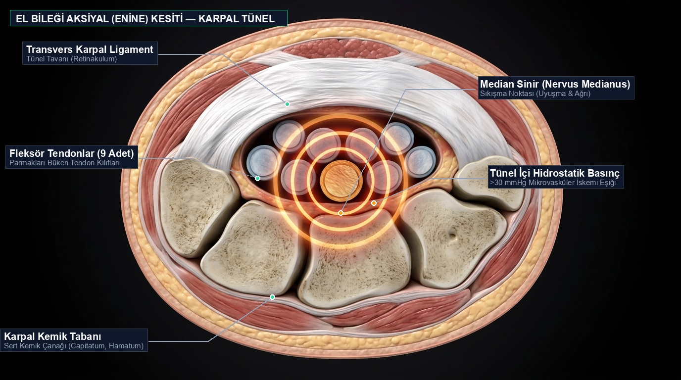 Figure 2: Anatomical transverse cross-section of the carpal tunnel, illustrating the transverse carpal ligament, nine flexor digitorum tendons, and median nerve vulnerable to microvascular ischemia at pressures exceeding 30 mmHg.
Intracarpal Hydrodynamic Pressure Model:
$P_{CT} = f(\theta_{\text{flex/ext}}, \theta_{\text{rad/ulnar}}, F_{\text{fingertip}}, F_{\text{grip}}, \text{External Contact Force})$
Clinical in vivo manometry reveals fundamental physiological baselines:
- Neutral Rest Baseline: In healthy individuals with wrists resting in neutral (approx. 2° extension, 2° ulnar deviation), intracarpal pressure is lowest: 2.5 to 8 mmHg [5, 7].
- Desk Contact Compression: Resting your wrists on a hard desk surface while typing or mousing spikes intracarpal canal pressure directly into the 30 to 55 mmHg range [8, 9].
- Keir’s Angular Safety Boundaries (2007): Keir et al. identified the angular thresholds that keep tunnel pressure under the critical 30 mmHg limit: ulnar deviation $\le 14.5^\circ$, radial deviation $\le 21.8^\circ$, extension $\le 32.7^\circ$, and flexion $\le 48.6^\circ$ [8].
- Microvascular Ischemic Threshold (30 mmHg): Epineural capillary perfusion pressure within the median nerve measures 30 to 40 mmHg. When intracarpal hydrostatic pressure exceeds 30 mmHg, capillary blood flow collapses, initiating epineural venous stasis, local hypoxia, axonal edema, and nocturnal paresthesia [6, 11].
3. Differential Diagnosis: Carpal Tunnel, De Quervain, and Lateral Tendinopathy
Repetitive Strain Injury (RSI) is an overarching diagnostic umbrella spanning three distinct clinical presentations common in screen and tool users [18]:
3.1. Carpal Tunnel Syndrome (CTS) — “Fingertip Paresthesia and Nocturnal Awakening”
Repeated tendon excursion under load causes tenosynovial hypertrophy. Werner & Andary (2011) showed that chronic mechanical strain thickens the transverse carpal ligament from its physiological 2 mm up to 4 mm, permanently restricting canal volume [12].
- Symptoms: Numbness, pins-and-needles, and burning in the thumb, index, middle finger, and radial half of the ring finger. Characteristic “flick sign” (shaking hands to restore blood flow upon waking).
3.2. De Quervain Tenosynovitis — “Smartphone Thumb”
Stenosing tenosynovitis of the first dorsal extensor compartment containing the abductor pollicis longus (APL) and extensor pollicis brevis (EPB) tendons.
- Etiology: Single-handed scrolling, typing, and reaching on phablet-sized displays forces the APL and EPB tendons to shear against the radial styloid process [13]. (Diagnostic confirmation via Finkelstein’s test: tuck the thumb into the palm, make a fist, and deviate the wrist ulnarly; sharp radial styloid pain indicates positive test).
- Anatomical Variants: Kulthanan & Chareonwat (2007) confirmed that individuals possessing an anatomical sub-compartmental septum separating APL and EPB have a markedly higher predisposition to De Quervain tenosynovitis under smartphone usage [15].
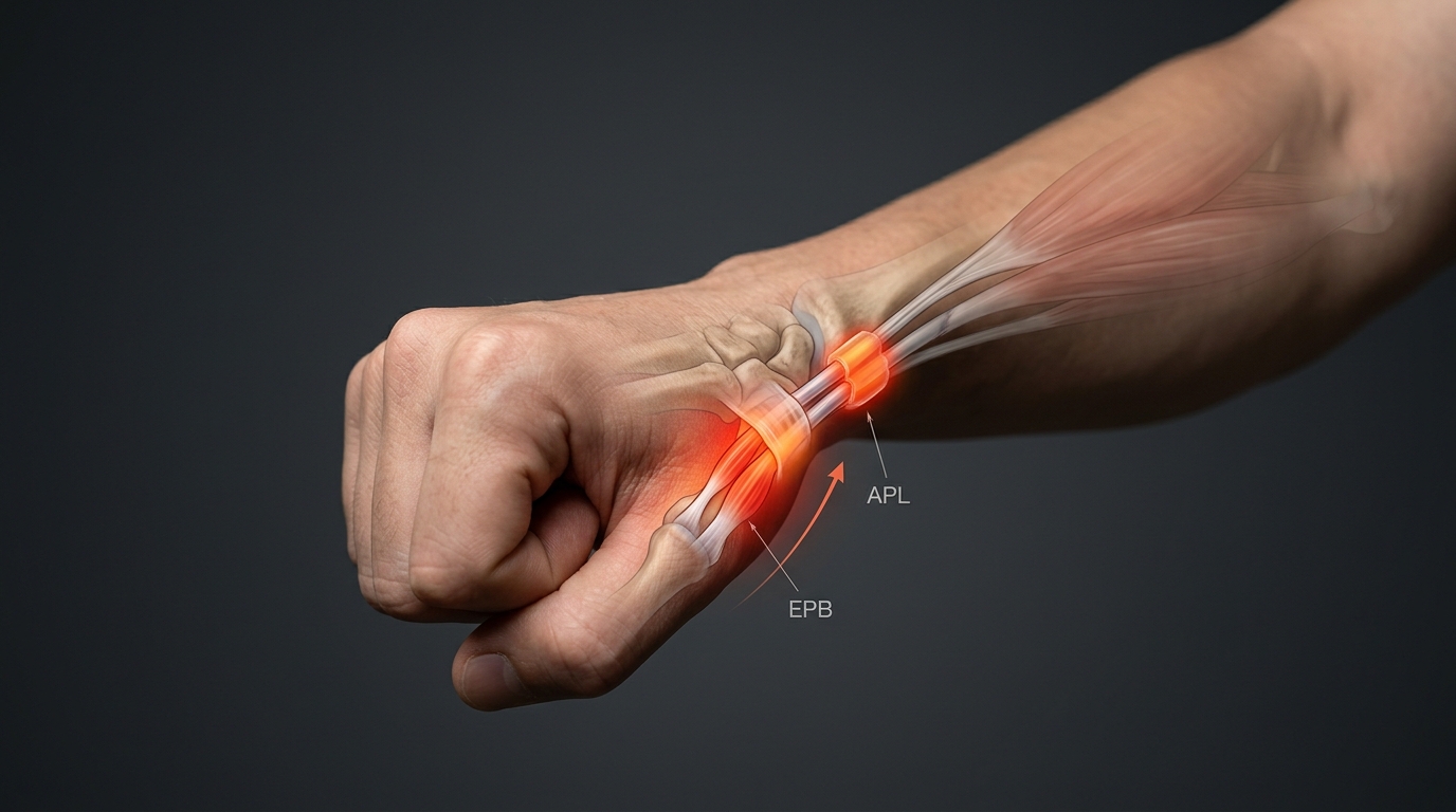 Figure 3: Friction of APL and EPB tendons within the first dorsal extensor compartment, De Quervain inflammatory changes, and anatomical rationale of Finkelstein’s test.
3.3. Lateral Elbow Tendinopathy — “Mouse Elbow / Tennis Elbow”
Sustained isometric extension of the wrist to elevate fingers over a keyboard or mouse engages the extensor carpi radialis brevis (ECRB) muscle continuously.
- Etiology: Conventional horizontal mice enforce 90° full forearm pronation, crossing the radius across the ulna and imposing chronic micro-avulsive strain at the common extensor origin on the lateral epicondyle [16].
4. Biomechanical Comparison Matrix: Everyday Devices & Ergonomic Solutions
| Device & Posture | Device Mass | Moment Arm & Angles | Primary Biomechanical Risk | Resulting Pathology | Ergonomic Solution & Alternative |
|---|---|---|---|---|---|
| Large Smartphone (One-Handed) | 200–240 g | Thumb reach (8–12 cm) | Extreme thumb abduction; pinky shelf base support | De Quervain tenosynovitis, basal thumb CMC joint osteoarthrosis | Two-handed typing, voice dictation, ring grip / PopSocket |
| Standard Flat Keyboard / Laptop | Fixed | Ulnar deviation (15°–25°), wrist on desk | Direct desk compression against carpal bones, hard keystrokes | Carpal tunnel syndrome ($P_{CT} > 30\text{ mmHg}$) | Gel palm support, negative-tilt split ergonomic keyboard |
| Standard Horizontal Mouse | 80–120 g | 90° full pronation | Wrist extension, carpal contact pressure, ECRB isometric loading | Lateral epicondylalgia (“mouse elbow”), median nerve ischemia | Vertical Ergonomic Mouse (57° natural handshake angle) |
| Industrial Mobile Computer / Scanner | 450–750 g | Forward-shifted CoM (12–18 cm) | High repetitive trigger force, long moment arm | Elevated moment impulse, flexor tenosynovitis | Glove-mounted barcode scanners, wearable ring terminals |
4.1. Deconstructing “Ergonomic Accessories”: Phone Grips and Vertical Mice
- Phone Grips (Rings & PopSockets): Rear-mounted rings eliminate the need to support the phone’s bottom edge with your pinky, alleviating “Smartphone Pinky” ligament strain. However, if you continue typing single-handed, thumb reaching torque remains high. The superior biomechanical method is two-handed operation or voice dictation.
- Why Vertical Mice Work: Resting your hand naturally on a flat surface orients the palm inward (neutral handshake position). Standard mice force 90° pronation, crossing the radius over the ulna and compressing the carpal canal floor. A vertical mouse restores a 57° neutral angle, reducing wrist and extensor tension by up to 70%.
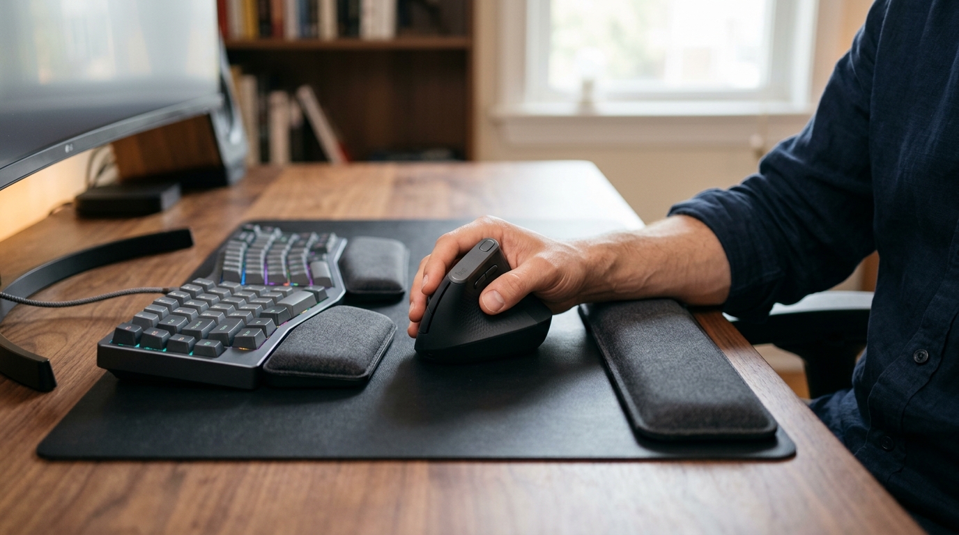 Figure 4: Vertical ergonomic mouse (57° neutral handshake angle) and split keyboard configuration preventing cross-over of forearm bones (radius-ulna).
5. Nutritional & Biochemical Protocols for Tendon and Nerve Health
Tendon extracellular matrix consists dry-weight of 60% to 85% collagen, of which ~95% is Type I collagen [19]. Vascularization in tendons is minimal, receiving only 5% to 10% of the blood supply of muscle tissue. Tenocytes depend on intermittent fluid perfusion driven by physical loading to absorb systemic nutrients [20].
[ TENDON BIOCHEMICAL REPAIR TIMING ]
45–60 Minutes Prior to Work/Loading:
15 g Hydrolyzed Collagen Peptides + 50–100 mg Vitamin C (Ascorbate)
                           │
                           ▼ (Mechanical Perfusion Coupling)
[ Pro-Hyp & Gly-Pro-Hyp Peak in Serum ]
                           │
                           ▼ (Prolyl Hydroxylase: Fe²⁺ Cofactor)
[ Stable Procollagen Triple-Helix Synthesis ] ──► [ Type I Collagen Fibrils & Cross-Links ]
5.1. Collagen Synthesis: Shaw Protocol (2017) and PINP Biomarker
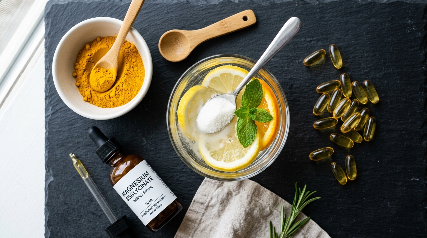 Figure 5: Tendon repair cascade and biochemical support: Hydrolyzed collagen peptides, vitamin C cofactor, magnesium bisglycinate, and anti-inflammatory omega-3 fatty acids.
- The Shaw Protocol: A landmark trial by Shaw et al. (2017) published in the American Journal of Clinical Nutrition proved that consuming 15 grams of vitamin C-enriched gelatin/collagen 45 to 60 minutes prior to intermittent mechanical loading doubles circulating levels of PINP (Procollagen I N-terminal Propeptide), the gold-standard biomarker for Type I collagen synthesis [21].
- Molecular Mechanism: Digested Pro-Hyp and Gly-Pro-Hyp tripeptides peak in serum at 60 minutes. Vitamin C (ascorbate) serves as an essential reducing cofactor, maintaining the $\text{Fe}^{2+}$ active center of prolyl hydroxylase. Without ascorbate, procollagen cannot fold into a stable triple helix [21, 22].
5.2. Magnesium Bisglycinate & Vitamin B6 (Toxicity Warning)
- Magnesium: Modulates NMDA receptor channels to dampen motor neuron hyperexcitability and enhance inhibitory chloride ($\text{Cl}^-$) influx in spinal cord motor neurons [23]. Tarsitano et al. (2024) demonstrated that correcting magnesium deficiencies aids muscle relaxation and reduces soreness [23].
- ⚠️ P-5-P and Sensory Neuropathy Risk: Pyridoxal 5-Phosphate (P-5-P) is an essential cofactor for glutamate decarboxylase in GABA synthesis [24]. However, a systematic review by Muhamad et al. (2023, Nutrients) cautioned that prolonged high-dose vitamin B6 supplementation (>50–100 mg/day) can trigger paradoxically disabling sensory peripheral neuropathy [25]. If burning, numbness, or tingling worsens, B6 intake must cease immediately.
5.3. Omega-3 Fatty Acids and Curcuminoids
- EPA & DHA: Compete with arachidonic acid for COX-2 enzymes, shifting downstream eicosanoid cascades from pro-inflammatory series-2 to anti-inflammatory series-3 prostaglandins ($\text{PGE}_3$) and specialized pro-resolving mediators (resolvins) [26, 27].
- Curcuminoids: Downregulate NF-$\kappa$B transcription and inhibit matrix metalloproteinases (MMPs) in inflamed synovial tissues [28, 29].
⚖️ Hierarchy of Interventions: 1. Biomechanical load reduction (shortening moment arms) ➔ 2. Graded therapeutic exercises ➔ 3. Clinical evaluation ➔ 4. Sleep & baseline nutrition ➔ 5. Targeted supplementation.
6. Circadian Biology, Nighttime Screen Exposure, and Tendon Healing
Late-night smartphone and computer use disrupts the central circadian pacemaker. In a meta-analysis of 33 studies by Wei et al. (2020), nocturnal light exposure was correlated with dramatic reductions in circulating melatonin metabolites [31].
[ Nighttime Screens & Blue Light (460 - 480 nm) ]
                     │
                     ▼
[ Intrinsically Photosensitive Retinal Ganglion Cells (ipRGCs) ]
                     │
                     ▼ Retinohypothalamic Tract (RHT)
[ Suprachiasmatic Nucleus (SCN) - Master Circadian Clock ]
                     │
        ┌────────────┴────────────┐
        ▼                         ▼
[ Pineal Gland Blocked ]    [ Nocturnal Cortisol Spikes ]
Melatonin Secretion CRASHES  Peaks between 02:00–04:00
        │                         │
        ▼                         ▼
* Tenocyte repair blunted        * Connective tissue inflammation persists
* Deep restorative sleep denied
6.1. Blue Light and Pineal Melatonin Suppression
Non-visual ipRGC cells in the retina, utilizing the photopigment melanopsin, are exquisitely sensitive to narrow-band blue light ($\lambda \approx 460 - 480\text{ nm}$) [32]. Light from phones and monitors stimulates ipRGCs, firing signals along the retinohypothalamic tract to the suprachiasmatic nucleus, tricking the brain into perceiving midday sunlight and suppressing pineal melatonin synthesis [33].
Melatonin is not merely a sleep-inducing molecule; it acts as a premier nocturnal antioxidant driving tenocyte proliferation and extracellular matrix remodeling during deep non-REM sleep [30]. Elevated nighttime cortisol impairs collagen synthesis, preventing daytime micro-fissures in tendon sheaths from healing [30].
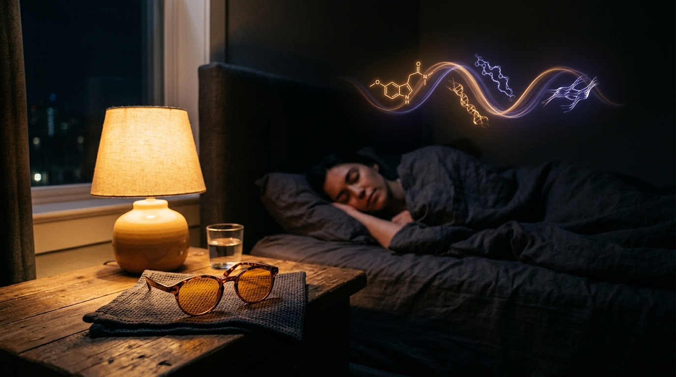 Figure 6: Photobiological pathway of blue light exposure, circadian melatonin downregulation, and impaired nighttime musculoskeletal collagen synthesis.
6.2. 3 Rules for Digital Workers and Night Shift Staff
- 1. Blue-Light Curfew 60 Minutes Pre-Bed: Discontinue handheld phone use in bed; activate hardware warm-amber color shifts or wear blue-attenuating eyewear [26].
- 2. Shift Worker Light Hygiene: Maintain bright lighting during active night shifts for alertness, but wear polarized dark sunglasses on the morning commute home to avoid resetting the central clock [34].
- 3. Sleep Sanctuary: Maintain a pitch-dark, cool sleeping environment (17–19°C / 63–66°F) to optimize slow-wave sleep and growth hormone (GH) secretion [36].
7. The 5-Minute Daily Wrist & Hand Maintenance Protocol
Following the “Sliders vs. Tensioners” neurodynamic principles established by Coppieters & Butler (2008), nerve mobilization should never stretch (tension) an irritable nerve; it must facilitate longitudinal gliding through surrounding soft-tissue interfaces [27].
[ 5-MINUTE DAILY WRIST & HAND MAINTENANCE PROTOCOL ]
┌───────────────┬────────────────────────────────────────────────────────┐
│ Minute 0 - 1  │ Baseline Check (Wrist, Thumb & Lateral Elbow Soreness) │
│ Minute 1 - 2  │ Ergonomic Reset (Keyboard Tilt, Mouse Angle, Phone)    │
│ Minute 2 - 3  │ 5-Stage Tendon Gliding (Straight Hand ➔ Full Fist)     │
│ Minute 3 - 4  │ Median & Radial Nerve Gliding (Neurodynamic Flossing)  │
│ Minute 4 - 5  │ Thenar Eminence (Basal Thumb) Myofascial Release       │
└───────────────┴────────────────────────────────────────────────────────┘- 0:00–1:00: Baseline Check: Assess subjective discomfort across the wrists, basal thumb joints, and lateral epicondyles.
- 1:00–2:00: Ergonomic Reset: Elevate wrists off hard surfaces; transition phone grip to two hands.
- 2:00–3:00: Five-Stage Tendon Gliding:
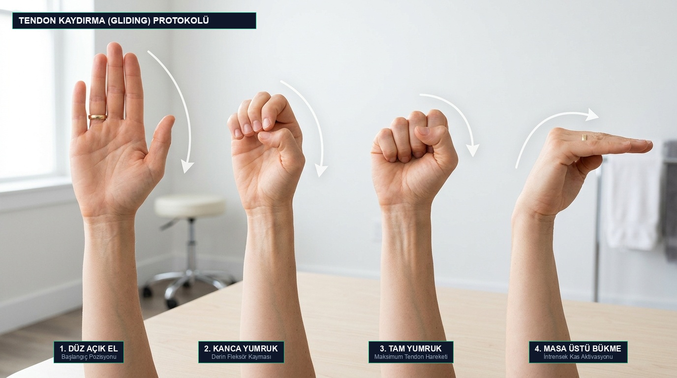 Figure 7: Four core tendon gliding positions for carpal tunnel health: Straight Open Hand, Hook Fist, Full Fist, and Tabletop Flat Bend.
Cycle smoothly through: (1) Straight open hand $\rightarrow$ (2) Hook fist $\rightarrow$ (3) Full fist $\rightarrow$ (4) Tabletop flat bend $\rightarrow$ (5) Straight fist. Hold each position for 2 to 3 seconds to ensure flexor tendons glide freely without adhesions [28]. 4. 3:00–4:00: Median Nerve Gliding (Neurodynamic Flossing): Abduct your arm laterally with wrist extended while gently tilting your head toward that shoulder. Then flex the wrist forward as you tilt your head away. Perform 10 fluid repetitions to mobilize the nerve without tension spikes [27]. 5. 4:00–5:00: Thenar Eminence Release: Apply gentle circular thumb pressure into the fleshy muscular base of your opposite thumb to release adductor pollicis and opponens pollicis spasm.
8. Actionable Daily Checklist
- Adopt Two-Handed Smartphone Operation: Replace single-handed thumb reaches with two-handed typing or voice dictation.
- Eliminate “Smartphone Pinky”: Stop resting the base weight of your phone onto your fifth digit; cradle the phone with your whole hand or use a desktop stand.
- Upgrade to a Vertical Mouse: For desk work exceeding 4 hours daily, switch to a 57° ergonomic vertical mouse to eliminate forearm bone crossing.
- Never Compress Wrists on Hard Desks: Float your wrists or rest palms on a soft gel support; maintain neutral carpal alignment.
- Use an Elevated Laptop Stand: Pair laptops with external split keyboards to prevent hyper-pronated, flexed wrist postures.
- Schedule 2-Minute Micro-Breaks Every 50 Minutes: Stand, perform tendon gliding sequences, and release thenar tension.
- Select Low-Trigger-Force Handhelds: In warehouse and industrial roles, deploy scanners with trigger actuation forces under 2 N and balanced centers of mass.
9. Frequently Asked Questions (FAQ)
Can Carpal Tunnel Syndrome be resolved without surgery?
Yes, conservative measures are frequently successful in mild to moderate stages. Wearing a neutral nocturnal wrist orthosis (0–5° extension), performing neurodynamic median nerve gliding drills, transitioning to vertical ergonomic mice, and adopting anti-inflammatory lifestyle strategies resolve symptoms for many patients. However, if thenar muscle atrophy or continuous sensory loss develops, prompt surgical decompression is mandatory to prevent permanent axonal degeneration.
Should I apply heat or cold (ice) to wrist pain?
Apply ice for acute flare-ups and tenosynovitis; use warmth for chronic muscular stiffness. Following intense typing or smartphone overuse, 10–15 minutes of cryotherapy helps constrict local capillaries and suppress intracarpal edema. For morning stiffness or forearm muscle tension, warm compresses stimulate local blood flow and tissue compliance.
When is the most critical time to wear a carpal tunnel splint?
During nocturnal sleep. When asleep, individuals instinctively flex their wrists, shooting intracarpal pressure over 30 mmHg and waking with numb hands. A rigid neutral nocturnal splint prevents flexion, allowing epineural microcirculation to recover overnight. Avoid wearing rigid splints continuously throughout the workday, as prolonged immobilization induces disuse muscle atrophy.
Is “Smartphone Pinky” a permanent bone deformity?
No, it is typically a transient soft-tissue indent, ligamentous strain, and muscular spasm. Resting heavy smartphones onto the distal interphalangeal or proximal interphalangeal joint of the little finger stresses the collateral ligaments and ulnar digital nerve. Switching to two-handed holding or utilizing phone grips resolves tenderness and soft-tissue swelling within a few weeks.
Which fingers are affected by Carpal Tunnel Syndrome, and why is the pinky spared?
Carpal tunnel syndrome affects only the thumb, index finger, middle finger, and the radial half of the ring finger. This specific distribution reflects the sensory innervation of the Median Nerve, which traverses the carpal canal. The pinky finger and the ulnar half of the ring finger are innervated by the Ulnar Nerve, which bypasses the carpal tunnel entirely via Guyon’s canal. If your pinky finger is numb, the compression is likely located at the elbow (cubital tunnel syndrome) or the cervical spine.
📚 Academic & Clinical References
- [1] National Institute for Occupational Safety and Health (NIOSH). (1997). Musculoskeletal Disorders and Workplace Factors: A Critical Review of Epidemiologic Evidence for Work-Related Musculoskeletal Disorders of the Neck, Upper Extremity, and Low Back. Cincinnati, OH: U.S. Department of Health and Human Services. DHHS (NIOSH) Publication No. 97-141.
- [2] Silverstein, B. A., Fine, L. J., & Armstrong, T. J. (1986). “Hand wrist cumulative trauma disorders in industry.” British Journal of Industrial Medicine, 43(11), 779–784. DOI: 10.1136/oem.43.11.779
- [3] Li, Z. M., Kuxhaus, L., Fisk, J. A., & Christophel, T. H. (2005). “Coupling between wrist flexion-extension and radial-ulnar deviation.” Clinical Biomechanics, 20(2), 177–183. DOI: 10.1016/j.clinbiomech.2004.10.002 • PubMed: 15621323
- [4] Schuind, F., Cooney, W. P., Linscheid, R. L., An, K. N., & Chao, E. Y. (1995). “Force and pressure transmission through the normal wrist. A theoretical two-dimensional biplanar model.” Journal of Biomechanics, 28(5), 587–601. DOI: 10.1016/0021-9290(94)00093-j • PubMed: 7775494
- [5] Weiss, N. D., Gordon, L., Bloom, T., So, Y., & Rempel, D. M. (1995). “Position of the wrist associated with the lowest carpal-tunnel pressure: implications for splint design.” The Journal of Bone and Joint Surgery. American Volume, 77(11), 1695–1699. DOI: 10.2106/00004623-199511000-00008 • PubMed: 7593062
- [6] Rempel, D., & Diao, E. (2004). “Entrapment neuropathies: pathophysiology and pathogenesis.” Journal of Orthopaedic Science, 9(2), 219–223. DOI: 10.1007/s00776-003-0744-y • PubMed: 15045543
- [7] Prayag, A. S., Najjar, R. P., & Gronfier, C. (2019). “Melatonin suppression is exquisitely sensitive to light and primarily driven by melanopsin in humans.” Journal of Pineal Research, 66(4), e12562. DOI: 10.1111/jpi.12562 • PubMed: 30697806
- [8] Boivin, D. B., & Boudreau, P. (2014). “Impacts of shift work on sleep and circadian rhythms.” Pathologie Biologie, 62(5), 292–301. DOI: 10.1016/j.patbio.2014.08.001 • PubMed: 25241979
- [9] Moore, J. S., & Garg, A. (1995). “The Strain Index: a proposed method to analyze jobs for risk of distal upper extremity disorders.” American Industrial Hygiene Association Journal, 56(5), 443–458. DOI: 10.1080/15428119591016863 • PubMed: 7604875
- [10] McGorry, R. W., Lin, J. H., & Dempsey, P. G. (2014). “Effect of grip type, wrist motion, and resistance level on pressures within the carpal tunnel of normal wrists.” Journal of Orthopaedic Research, 32(5), 697–705. DOI: 10.1002/jor.22571 • PubMed: 24391059
- [11] Gelberman, R. H., Hergenroeder, P. T., Hargens, A. R., Lundborg, G. N., & Akeson, W. H. (1981). “The carpal tunnel syndrome. A study of carpal canal pressures.” The Journal of Bone and Joint Surgery. American Volume, 63(3), 380–383. PubMed: 7204435
- [12] Werner, R. A., & Andary, M. (2011). “Electrodiagnostic evaluation of carpal tunnel syndrome.” Muscle & Nerve, 44(4), 597–607. DOI: 10.1002/mus.22208 • PubMed: 21915874
- [13] Fakoya, A. O., & Oluwadiya, K. S. (2023). “De Quervain’s Disease: A Discourse on Etiology, Diagnosis, and Treatment.” Cureus, 15(4), e38079. DOI: 10.7759/cureus.38079 • PubMed: 37252462
- [14] Stahl, S., Vida, D., Meisner, C., Lotter, O., Rothenberger, J., Schaller, H. E., & Stahl, A. S. (2013). “Systematic review and meta-analysis on the work-related cause of de Quervain tenosynovitis: a critical appraisal of its recognition as an occupational disease.” Plastic and Reconstructive Surgery, 132(6), 1479–1491. DOI: 10.1097/01.prs.0000434409.32594.1b • PubMed: 24005369
- [15] Kulthanan, T., & Chareonwat, B. (2007). “Variations in abductor pollicis longus and extensor pollicis brevis tendons in the Quervain syndrome.” Scandinavian Journal of Plastic and Reconstructive Surgery and Hand Surgery, 41(1), 36–38. DOI: 10.1080/02844310600869720 • PubMed: 17484184
- [16] Kraushaar, B. S., & Nirschl, R. P. (1999). “Tendinosis of the elbow (tennis elbow). Clinical features and findings of histological, immunohistochemical, and electron microscopy studies.” The Journal of Bone and Joint Surgery. American Volume, 81(2), 259–278. PubMed: 10073590
- [17] Curti, S., Mattioli, S., Bonfiglioli, R., Farioli, A., & Violante, F. S. (2021). “Elbow tendinopathy and occupational biomechanical overload: a systematic review with best-evidence synthesis.” Journal of Occupational Health, 63(1), e12186. DOI: 10.1002/1348-9585.12186 • PubMed: 33534951
- [18] van Tulder, M., Malmivaara, A., & Koes, B. (2007). “Repetitive strain injury.” The Lancet, 369(9575), 1815–1822. DOI: 10.1016/S0140-6736(07)60820-4 • PubMed: 17531890
- [19] Screen, H. R., Berk, D. E., Kadler, K. E., Ramirez, F., & Young, M. F. (2015). “Tendon functional extracellular matrix.” Journal of Orthopaedic Research, 33(6), 793–799. DOI: 10.1002/jor.22818 • PubMed: 25640030
- [20] Kjaer, M., Langberg, H., Heinemeier, K., et al. (2009). “From mechanical loading to collagen synthesis, structural changes and function in human tendon.” Scandinavian Journal of Medicine & Science in Sports, 19(4), 500–510. DOI: 10.1111/j.1600-0838.2009.00986.x • PubMed: 19706001
- [21] Shaw, G., Lee-Barthel, A., Ross, M. L., Wang, B., & Baar, K. (2017). “Vitamin C-enriched gelatin supplementation before intermittent activity augments collagen synthesis.” The American Journal of Clinical Nutrition, 105(1), 136–143. DOI: 10.3945/ajcn.116.138594 • PubMed: 27852613
- [22] Hijlkema, A., Roozenboom, C., Mensink, M., & Zwerver, J. (2022). “The impact of nutrition on tendon health and tendinopathy: a systematic review.” Journal of the International Society of Sports Nutrition, 19(1), 474–504. DOI: 10.1080/15502783.2022.2104130 • PubMed: 35937777
- [23] Tarsitano, M. G., et al. (2024). “Effects of magnesium supplementation on muscle soreness in different type of physical activities: a systematic review.” Journal of Translational Medicine, 22(1), 609. DOI: 10.1186/s12967-024-05434-x • PubMed: 38970118
- [24] Grootswagers, P., et al. (2021). “Vitamin B-6 intake is related to physical performance in European older adults: Results from the NU-AGE study.” The American Journal of Clinical Nutrition, 113(4), 840–848. DOI: 10.1093/ajcn/nqaa368 • PubMed: 33515034
- [25] Muhamad, R., et al. (2023). “The Role of Vitamin B6 in Peripheral Neuropathy: A Systematic Review.” Nutrients, 15(13), 2823. DOI: 10.3390/nu15132823 • PubMed: 37447150
- [26] Sandford, F. M., Sanders, T. A., & Lewis, J. S. (2018). “A randomised controlled trial of long-chain omega-3 polyunsaturated fatty acids in rotator cuff related shoulder pain.” BMJ Open Sport & Exercise Medicine, 4(1), e000414. DOI: 10.1136/bmjsem-2018-000414 • PubMed: 30364577
- [27] Fernández-Lázaro, D., et al. (2024). “Omega-3 supplementation on post-exercise inflammation, muscle damage, oxidative response and sports performance: a systematic review.” Nutrients, 16(13), 2044. DOI: 10.3390/nu16132044 • PubMed: 38999792
- [28] Córdova, A., et al. (2023). “Is Curcumine Useful in the Treatment and Prevention of Tendinopathy and Myotendinous Junction Injury?” Nutrients, 15(2), 384. DOI: 10.3390/nu15020384 • PubMed: 36678255
- [29] Oxley, J., & Peart, D. J. (2024). “Curcumin supplementation and exercise-induced muscle damage: a systematic review and meta-analysis.” Nutrition and Health, 30(2), 177–187. DOI: 10.1177/02601060231186439 • PubMed: 37408367
- [30] Boivin, D. B., Boudreau, P., & Kosmadopoulos, A. (2022). “Disturbance of the Circadian System in Shift Work and Its Health Impact.” Journal of Biological Rhythms, 37(1), 3–28. DOI: 10.1177/07487304211064218 • PubMed: 34969316
- [31] Wei, T., Li, C., Heng, Y., et al. (2020). “Association between night-shift work and level of melatonin: systematic review and meta-analysis.” Sleep Medicine, 75, 502–509. DOI: 10.1016/j.sleep.2020.09.018 • PubMed: 33022488
- [32] Mure, L. S. (2021). “Intrinsically photosensitive retinal ganglion cells of the human retina.” Frontiers in Neurology, 12, 636330. DOI: 10.3389/fneur.2021.636330 • PubMed: 33841306
- [33] Vartanian, G. V., et al. (2015). “Melatonin suppression by light in humans is more sensitive than previously reported.” Journal of Biological Rhythms, 30(4), 351–354. DOI: 10.1177/0748730415585413 • PubMed: 26017927
- [34] National Institute for Occupational Safety and Health (NIOSH). (2020). NIOSH Training for Nurses on Shift Work and Long Work Hours: Module 9 - Light and Sleep Management. Cincinnati, OH: CDC/NIOSH.
- [35] Keir, P. J., Bach, J. M., Hudes, M., & Rempel, D. M. (2007). “Guidelines for wrist posture based on carpal tunnel pressure thresholds.” Human Factors, 49(1), 128–139. DOI: 10.1518/001872007779598127 • PubMed: 17315846
- [36] Lam, C., & Chung, M. H. (2021). “Dose-response effects of light therapy on sleepiness and circadian phase shift in shift workers: a meta-analysis and moderator analysis.” Scientific Reports, 11(1), 11560. DOI: 10.1038/s41598-021-89321-1 • PubMed: 34099750
For informational purposes only; consult a physician for medical diagnosis or treatment.