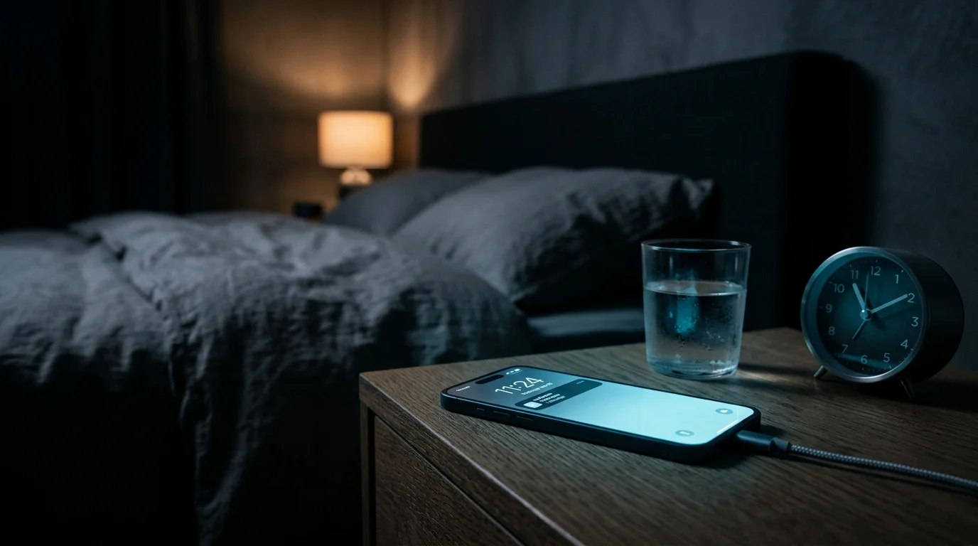
- → Your Eyes Tell Time, Not Just Vision: Retinal ganglion layers contain specialized non-visual photoreceptors called intrinsically photosensitive retinal ganglion cells (ipRGCs) powered by the photopigment melanopsin.
- → Deceiving the Central Pacemaker: When ipRGCs capture blue photons (~480 nm) emitted by smartphones, they dispatch an immediate 'daytime' signal to the suprachiasmatic nucleus (SCN).
- → Suppression of Dim Light Melatonin Onset (DLMO): Photic stimulation of the SCN triggers sympathetic inhibition of the pineal gland, acutely suppressing and delaying nocturnal melatonin synthesis.
- → Tired But Wired Paradox: Even when adenosine pressure (Process S) is elevated from a long day, photic circadian phase shifting (Process C) flips the nervous system into physiological hyperarousal.
- → Dark Room Contrast Hazard: Staring at a solitary luminous screen in an unlit bedroom concentrates lux density onto the fovea and parafovea, multiplying melanopic suppression.
🧭 Circadian Light Hygiene Series (Part 1/3):
📍 Part 1 (Current Guide): Nighttime Smartphone Use and the Retinal Circadian Switch (ipRGCs)
⏭️ Part 2: Do Blue Light Glasses and Night Shift Really Work? 2026 Clinical Evidence
⏭️ Part 3: Morning Photic Anchoring: Setting the Central Clock with Early Sunlight
⚠️ Medical Disclaimer: This guide is intended solely for scientific education, sleep architecture optimization, and photobiological awareness. It does not replace clinical consultation, medical diagnosis, or targeted insomnia treatment. If you suffer from chronic insomnia or circadian rhythm sleep disorders, seek evaluation from a board-certified sleep specialist.
🟢 Evidentiary Level: Systematic Review & Clinical Trials — Grounded in human trials evaluating objective circadian biomarkers (DLMO, salivary/plasma melatonin kinetics, polysomnography).
What This Guide Explores
We have all experienced this familiar evening scenario: After an exhausting workday, your eyelids grow heavy and you sink into bed, eager to sleep. Yet, out of habit, you reach for your phone for “just five minutes to check notifications.”
Moments later, an unsettling biological shift occurs: The heavy wave of sleepiness evaporates into thin air. Even after putting the device down, you toss and turn, feeling alert, restless, and unable to drift off.
Most people blame the cognitive excitement or dopamine hits of social media feeds. Psychological arousal certainly plays a role; however, the primary, non-negotiable culprit is an ancient, mechanical photobiological reflex.
Drawing upon a landmark 2026 systematic review of human circadian studies [1], this guide breaks down how the hidden circadian switch behind your retinas operates, and outlines practical steps to protect your sleep architecture without abandoning modern technology.
1. The Retinal Master Switch: Intrinsically Photosensitive Retinal Ganglion Cells (ipRGCs)
Classical sensory physiology historically identified two types of retinal photoreceptors: cones (mediating daylight visual acuity and color perception) and rods (mediating low-light scotopic vision).
However, in the early 2000s, neuroscientists discovered a third, non-image-forming photoreceptor class: intrinsically photosensitive retinal ganglion cells (ipRGCs) [2].
These specialized neurons do not construct visual imagery. Instead, their evolutionary purpose is singular: to gauge environmental irradiance and synchronize the body’s internal master clock to the planetary solar cycle.
ipRGCs express the photopigment melanopsin. Axons from these cells bypass the visual cortex entirely and travel along the retinohypothalamic tract (RHT) straight to the brain's circadian pacemaker — the Suprachiasmatic Nucleus (SCN). The instant evening photons stimulate this circuit, the SCN deduces that it is midday, slamming the brakes on sleep-promoting hormone cascades.
2. Why Your Brain Mistakes a Smartphone Screen for Noon Sunlight
Melanopsin does not respond uniformly to all wavelengths. Its spectral sensitivity curve exhibits a sharp peak at ~480 nanometers (nm)—the characteristic narrow band of high-energy visible cyan-blue light.
In nature, this wavelength dominates the midday sky on a cloudless afternoon.
- Modern smartphone, tablet, and LED displays emit peak spectral irradiance precisely in this ~450–480 nm band.
- When you look at an emissive display in a dark bedroom, these photons strike melanopsin-bearing ipRGCs, firing relentless action potentials into the hypothalamus.
A comprehensive 2026 systematic review [1] confirmed that inappropriate nocturnal stimulation of human ipRGCs directly postpones Dim Light Melatonin Onset (DLMO), blunting nighttime plasma melatonin peaks and fracturing circadian entrainment.
As specialist Marianne Krug highlighted in clinical discussions on chronobiology [5], melatonin is not merely a mild sedative, but an endocrine master metronome. When high nocturnal light exposure elevates sympathovagal tone and keeps cortisol unseasonably high, natural melatonin synthesis is paralyzed. Consequently, even if physical exhaustion allows someone to fall asleep, they frequently awaken abruptly between 11:00 PM and 3:00 AM, suffering from fragmented sleep architecture (Durchschlafstörungen).

3. The “Tired But Wired” Neurobiology
In Alexander Borbély’s foundational Two-Process Model of Sleep Regulation (1982) [4], healthy slumber depends on the harmonious alignment of two distinct forces:
- Process S (Homeostatic Sleep Drive): Driven by the progressive accumulation of metabolic waste products, primarily adenosine, across hours of wakefulness.
- Process C (Circadian Drive): An oscillatory rhythmic clock governed by the SCN and synchronized by light.
When you slide beneath the covers after a grueling 16-hour day, Process S is peaking; your neurons are saturated with adenosine. Your body is biologically primed to sleep. However, the moment your eyes lock onto an illuminated phone screen, Process C is forced in the opposite direction.
While peripheral tissues cry out for cellular repair, the central clock proclaims “It is broad daylight!”, maintaining paraventricular suppression over the pineal gland. This biochemical clash induces the classic “tired but wired” neurostate: deep muscular exhaustion paired with hyperalert cerebral agitation.
Furthermore, as Dr. Kurt Mosetter emphasizes in cellular sleep medicine [6], late-night exposure to blue-shifted light downregulates core somatic “clock genes” (Schlaf-Gene). Falling asleep while passively staring at a streaming video or phone display does not yield restorative sleep; it flattens slow-wave deep sleep and disrupts REM stages, leaving you groggy and unrefreshed the next morning.
4. The Practical Light Hygiene Protocol
Overcoming nocturnal insomnia does not require discarding your digital devices; it requires aligning display technology with your eye’s photobiological sensitivity curve.
Step 1: Transition to Warm Ambient Dimness
- At least two hours before sleep, extinguish overhead cool-white (5000K–6500K) LED fixtures.
- Rely on low-positioned, warm incandescent bulbs, amber lamps (<2700K), or floor lighting positioned below eye level.
Step 2: Never Use Screens in Pitch Darkness
Viewing an unbuffered, luminous display in an otherwise pitch-black room creates an extreme contrast ratio. The pupillary aperture dilates in the dark periphery, exposing the central retina to concentrated melanopic lux.
- Always ensure soft, diffuse background lighting if late-night digital work is unavoidable.
- Lower display luminance to its absolute lowest hardware threshold.
Step 3: Enforce a 60-Minute Photic Buffer Before Sleep
Establish an unbreakable one-hour screen curfew prior to closing your eyes.
- Deprived of direct blue-cyan photons, the SCN releases its brake on the pineal gland, allowing DLMO to surge unimpeded.
- Use this 60-minute wind-down window to lower autonomic tone with 0.1 Hz resonance breathing, or read an analog paper book or reflection-based E-Ink device.
5. Common Misconceptions to Avoid
- “Night Shift / True Tone Is Active, So I Can Scroll Safely”:
Warming software colors shifts spectral output toward amber, but does not eradicate total melanopic irradiance. At high brightness levels in a pitch-black bedroom, software color shifts alone still activate ipRGCs and delay DLMO. - “Standard Clear Blue Light Glasses Shield My Sleep”:
Over-the-counter clear computer glasses typically filter only 10% to 20% of high-energy blue photons. To fully shield ipRGC melanopsin from nocturnal light, amber- or red-tinted lenses blocking 98%+ of wavelengths below 500 nm are required.
6. High-Risk Populations and Contraindications
- Ophthalmic Conditions (Retinopathy & Glaucoma): As highlighted in the 2026 review [1], individuals with advanced retinal degenerative disease or open-angle glaucoma experience altered ipRGC density, which can cause severe autonomous circadian misalignment.
- Psychological Revenge Bedtime Procrastination: Light exposure interacts powerfully with behavioral delays. When smartphone use becomes an emotional coping mechanism to reclaim autonomy, photobiological interventions must be paired with behavioral cognitive boundary-setting.
7. Verified Methodology & Scientific References
- Intrinsically Photosensitive Retinal Ganglion Cell Dysfunction and Circadian Disruption (2026):
Intrinsically Photosensitive Retinal Ganglion Cell Dysfunction and Suprachiasmatic Nucleus-Mediated Circadian Disruption in Humans: A Systematic Review. Experimental Eye Research.
PMID: 42692181 - Melanopsin and Circadian Phototransduction (2002):
Berson, D. M., Dunn, F. A., & Takao, M. Phototransduction by retinal ganglion cells that set the circadian clock. Science, 295(5557), 1070-1073.
PMID: 11834835 - Evening Room Light and Melatonin Suppression (2011):
Gooley, J. J., et al. Exposure to room light before bedtime suppresses melatonin onset and shortens melatonin duration in humans. J Clin Endocrinol Metab, 96(3), E463-E472.
PMID: 21193540 - The Two-Process Model of Sleep Regulation (1982):
Borbély, A. A. A two-process model of sleep regulation. Human Neurobiology, 1(3), 195-204.
PMID: 7185792 - Cortisol Dysregulation, Sleep Continuity, and Melatonin Rhythm:
Marianne Krug. Das Durchschlafproblem und das wahre Schlafhormon. QS24 Swiss Health Television. YouTube. - Disrupted Melatonin Dynamics, Blue Light, and Sleep Architecture:
Dr. Kurt Mosetter & Henning Fritz. Gestörter Melatonin-Haushalt. QS24 Swiss Health Television. YouTube.
For informational purposes only; consult a physician for medical diagnosis or treatment.