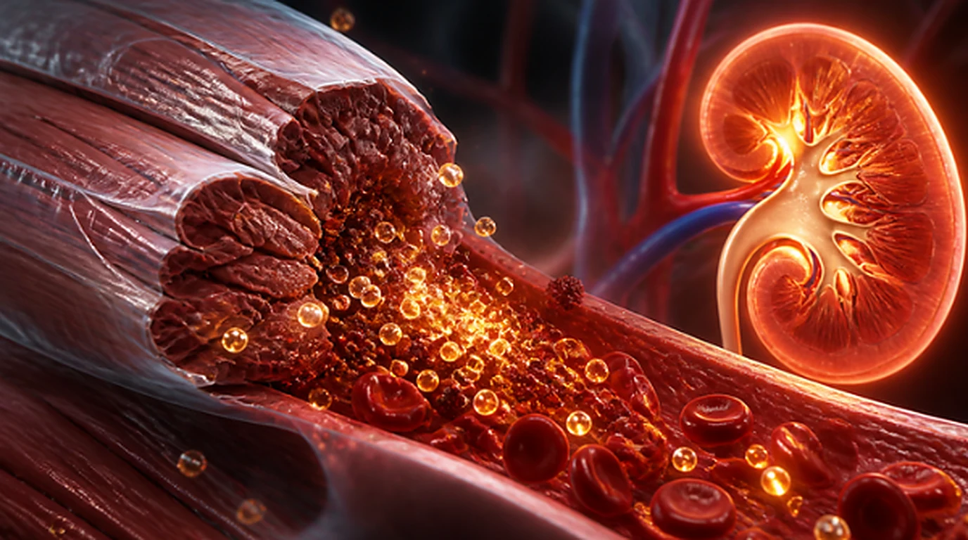
- → Bywaters Syndrome and the Muscle Electrolyte Pool (Bywaters & Beall, 1941): Skeletal muscle comprises ~40% of adult body mass and harbors over 70% of total body potassium (K⁺); when the sarcolemma ruptures, this massive reservoir floods directly into the bloodstream.
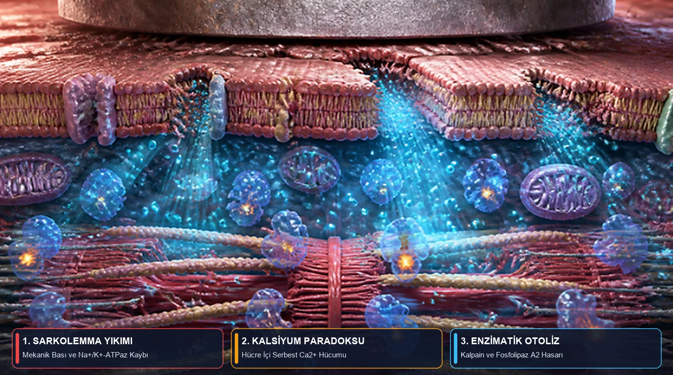
- → The Intracellular Calcium (Ca²⁺) Paradox (Bosch, Poch, & Grau, 2009): Mechanical compression or ATP depletion impairs ion pumps, elevating cytosolic free Ca²⁺ up to 100-fold and activating neutral proteases (calpains) and phospholipases that digest the muscle cell from within.
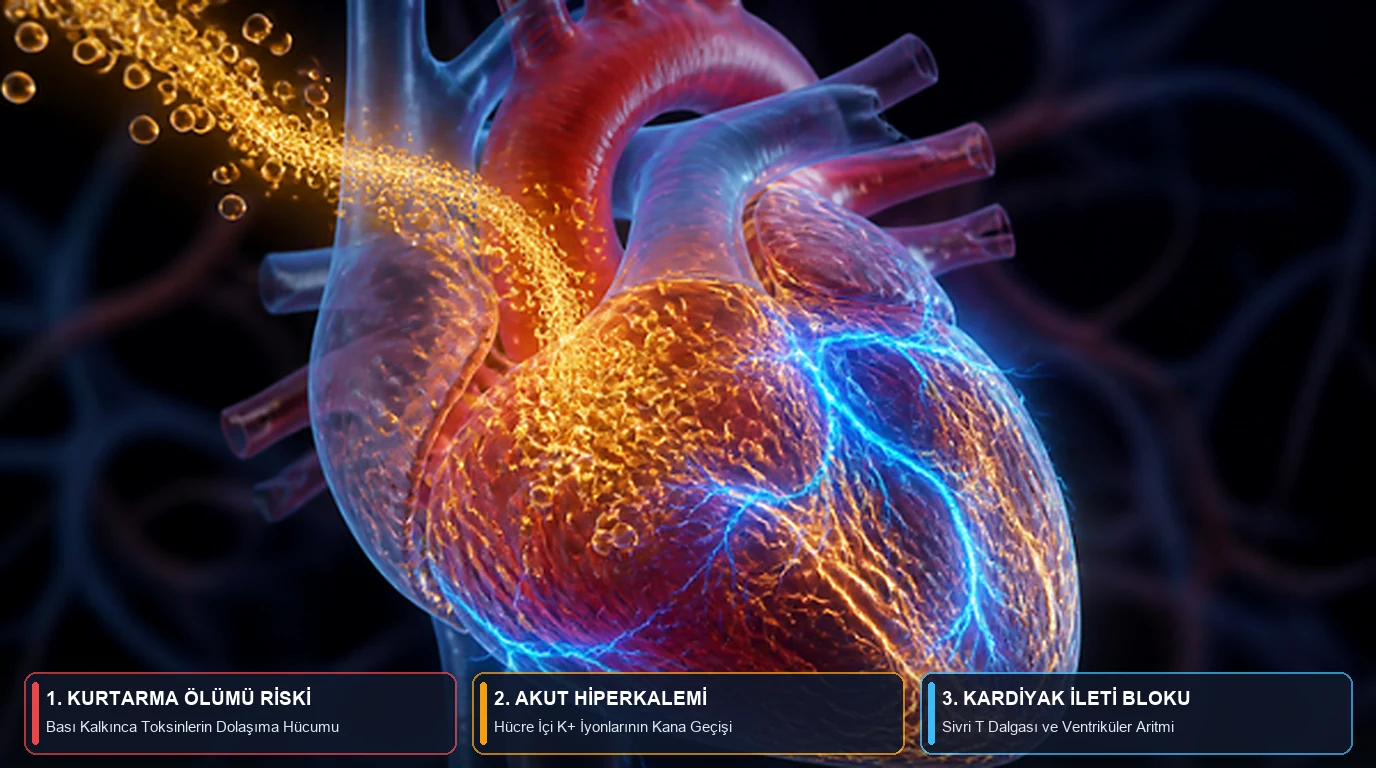
- → Reperfusion and 'Rescue Death' (Sever, Vanholder, & Lameire, 2006): Decompressing an entrapped limb triggers rapid hypovolemia as fluid sequestrates into swollen muscle ('third spacing'), while surging extracellular potassium (>6.5 mmol/L) collapses cardiac resting potentials, precipitating fatal ventricular arrhythmias.
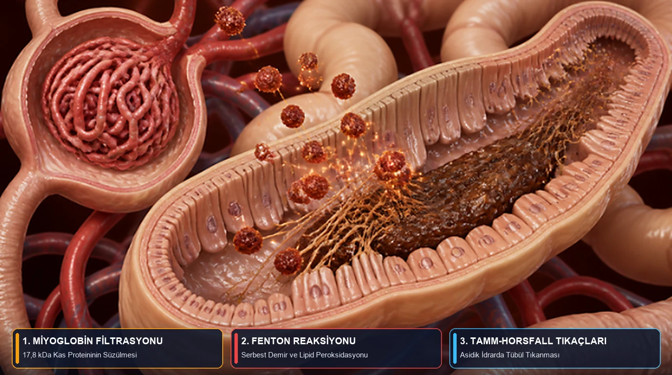
- → Myoglobinuria and Nephrotoxicity: The released 17.8 kDa monomeric myoglobin induces Acute Kidney Injury (AKI) through three converging pathways: renal vasoconstriction via nitric oxide scavenging, heme-iron (Fe²⁺/Fe³⁺) Fenton-mediated lipid peroxidation, and acidic intratubular cast formation with Tamm-Horsfall protein.
⚠️ Emergency Medical Disclaimer: The information presented in this guide is derived from published literature in muscle physiology, nephrology, and disaster trauma medicine. It is intended strictly for scientific education and cannot replace professional medical judgment, paramedic protocols, or formal emergency first aid training. If an individual experiences entrapment, severe crush trauma, or develops tea/cola-colored dark urine, acute muscle swelling, severe oliguria, or cardiac palpitations following intense exertion, contact emergency services (911 / 112) immediately.
Executive Summary
- Core Definition: Rhabdomyolysis is the rapid dissolution of damaged skeletal muscle fibers resulting in the release of intracellular contents (potassium
K⁺, creatine kinaseCK, myoglobin, phosphate, urate) into systemic circulation; its systemic traumatic manifestation following prolonged compression is known as Crush Syndrome [1], [2], [3]. - The Calcium Cascade: When mechanical ischemia depletes ATP, membrane
Na⁺/K⁺-ATPaseandCa²⁺-ATPasepumps stall. Cytosolic freeCa²⁺skyrockets to 100 times baseline, locking myofilaments into irreversible contracture and activating self-destructive lytic enzymes [1]. - Cardiac Threat (Hyperkalemia): Skeletal myocytes contain
~140 mmol/Lof potassium, contrasted against a narrow plasma range of3.5–5.0 mmol/L. Rapid dumping of this pool into venous circulation provokes lethal hyperkalemic cardiac arrest within minutes of extrication [2], [3]. - Renal Threat (Myoglobinuria): Monomeric myoglobin filters freely through the glomerulus, coloring urine dark reddish-brown (“tea or cola color”) and provoking Acute Kidney Injury (AKI) via oxidative tubular damage and luminal cast obstruction under acidic conditions [1], [3], [4].
Introduction: Why Does Extrication Trigger Fatal Collapse?
In structural collapses, industrial machinery accidents, or natural disasters, emergency responders frequently witness one of the most sobering paradoxes in trauma medicine: a pinned survivor may remain conscious, stable, and conversational while pinned beneath a heavy beam. Yet, within minutes or hours after the compressing weight is lifted, the patient may experience sudden cardiac arrest (rescue death) or develop anuric renal failure over the subsequent 48 hours [2], [3].
First formally documented during the 1941 London Blitz by Dr. Eric Bywaters and Desmond Beall in the British Medical Journal (BMJ), this entity—termed Bywaters Syndrome (Crush Syndrome)—demonstrates how localized ischemic muscle trauma translates into catastrophic systemic metabolic shock [2]. Beyond structural disasters, rhabdomyolysis frequently emerges in civilian life: unaccustomed exhaustive eccentric exertion in hot environments (exertional rhabdomyolysis), prolonged immobilization following toxic overdoses, or statin-drug interactions [1], [3].
1. The Cellular Breakdown: Sarcolemmal Rupture and the Calcium (Ca²⁺) Cascade
Striated skeletal muscle represents the single largest tissue mass in the human body (accounting for roughly 40% of adult body weight). In healthy myocytes, continuous ATP-driven transport through sarcolemmal ion pumps maintains steep electrochemical gradients: high intracellular potassium (K⁺ ≈ 140 mmol/L) and negligible cytosolic calcium (Ca²⁺ ≈ 0.1 µmol/L), opposed by high extracellular sodium (Na⁺ ≈ 140 mmol/L) and calcium (Ca²⁺ ≈ 1–2 mmol/L) [1], [3].
As synthesized by Xavier Bosch, Esteban Poch, and Josep M. Grau in The New England Journal of Medicine (NEJM), regardless of whether the initiating insult is blunt mechanical crushing or metabolic substrate starvation, rhabdomyolysis converges upon a final common pathway: uncontrolled accumulation of free cytosolic calcium (Ca²⁺) [1]:
- Mechanical Shear and Ischemic ATP Depletion: Sustained mechanical compression shears the sarcolemma, producing direct micro-tears. Simultaneously, localized vascular occlusion deprives tissue of oxygen, rapidly depleting intracellular ATP reserves [1], [3].
- Ion Transporter Failure: Lacking ATP, both the
Na⁺/K⁺-ATPaseand the sarcoplasmic/sarcolemmalCa²⁺-ATPasefail. Intracellular sodium rises, pulling in water and causing marked myocyte edema; concurrently, theNa⁺/Ca²⁺exchanger operates in reverse mode, flooding the cytosol with extracellularCa²⁺[1]. - Calpain and Phospholipase Activation: Sustained cytosolic
Ca²⁺elevations (reaching 10- to 100-fold normal resting levels) initiate irreversible enzymatic degradation:- Actin-myosin cross-bridges lock into persistent, ATP-depleting contraction [1].
- Calcium-dependent neutral proteases (calpains) and phospholipase A₂ (
PLA₂) activate, dismantling myofibrillar structural proteins, mitochondrial membranes, and sarcolemmal phospholipids [1], [3]. - Mitochondria swell under excessive calcium sequestration, generating massive reactive oxygen species (ROS) and consummating myocyte necrosis [1].
2. The Reperfusion Surge and “Rescue Death”: How Hyperkalemia Paralyzes the Myocardium
Pioneering investigations by Prof. Mehmet Şükrü Sever (Istanbul University) and Prof. Raymond Vanholder (Ghent University) in The New England Journal of Medicine (NEJM) and JASN delineate the violent, bidirectional shockwave that occurs the moment compressive force is relieved (decompression / reperfusion) [3], [4]:
2.1. “Third-Space” Fluid Sequestration and Hypovolemic Shock
When perfusion is restored to necrotic, permeabilized limb musculature, capillary hydrostatic pressure drives massive fluid transudation. Liters of plasma water, albumin, and sodium rush out of the vascular tree into disrupted muscle beds (third-space fluid sequestration). An entrapped thigh or calf can sequester 4 to 6 liters of extracellular fluid within hours, leading to precipitous hypovolemic shock and profound renal hypoperfusion [3], [4].
2.2. The Potassium (K⁺) Tsunami and Cardiac Electrophysiology
Simultaneously, the release of venous obstruction unleashes a flood of necrotic intracellular metabolites into the central venous circulation [1], [3], [4]:
| Released Cellular Constituent | Biochemical Consequence | Target Organ & Primary Clinical Risk |
|---|---|---|
1. Potassium (K⁺) | Drives serum K⁺ rapidly past 6.0–7.0 mmol/L (Hyperkalemia). | Myocardium: Collapses resting membrane potential; generates tall, peaked T waves, PR prolongation, and QRS widening, culminating in ventricular fibrillation and asystole [3, 4]. |
2. Phosphate (PO₄³⁻) | Induces marked Hyperphosphatemia, binding ionized calcium into ectopic micro-calcifications. | Cardiovascular & Neuromuscular: Triggers acute Hypocalcemia, which synergistically exacerbates potassium-induced myocardial arrhythmogenicity [1, 3]. |
| 3. Lactic and Organic Acids | Flushes ischemic metabolites into systemic circulation, inducing High Anion Gap Metabolic Acidosis. | Systemic Vasculature & Kidneys: Acidemia promotes further cellular K⁺ efflux and accelerates intratubular myoglobin precipitation [1, 3]. |
4. Myoglobin (17.8 kDa) | Exceeds serum binding capacity (haptoglobin) and filters freely across the glomerular basement membrane (Myoglobinuria). | Renal Tubules: Drives Acute Kidney Injury (AKI) via vasoconstriction, lipid peroxidation, and tubular cast obstruction [1, 4]. |
5. Creatine Kinase (CK) | Serum CK (normally < 200 U/L) escalates into the range of 5,000 to >100,000 U/L. | Diagnostic Biomarker: Gold-standard surrogate marker reflecting the quantitative magnitude of skeletal muscle necrosis [1, 3]. |
3. How Myoglobinuria Sabotages Renal Function: The Triad of Nephrotoxicity
Myoglobin is an intracellular hemoprotein responsible for oxygen storage in striated muscle. Unlike the tetrameric 64.5 kDa hemoglobin molecule, myoglobin exists as a compact 17.8 kDa single-chain monomer [1], [4]. Because of this small molecular weight and weak plasma protein binding, once serum concentrations exceed 0.5–1.5 mg/dL, myoglobin saturates haptoglobin and passes readily through glomerular fenestrations. Upon reaching urinary thresholds of ~100 mg/dL, it imparts a distinct tea-, cola-, or reddish-brown coloration to the urine [1], [3].
Comprehensive work by Bosch et al. (NEJM, 2009) and Vanholder et al. (JASN, 2000) demonstrates that myoglobin destroys nephrons via three concurrent, synergistic mechanisms [1], [4]:
- Renal Vasoconstriction: Third-spacing hypovolemia drives powerful neurohumoral vasoconstriction (sympathetic tone, endothelin-1, and angiotensin II). Simultaneously, freely filtered myoglobin actively scavenges endothelial nitric oxide (
NO), abolishing intrarenal vasodilation and inducing profound medullary ischemia [1], [4]. - Heme-Iron (
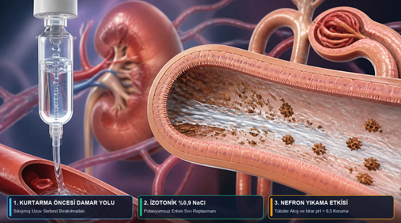
Fe²⁺ → Fe³⁺) Redox Cycling & Fenton Reaction: Endocytosed by proximal tubular epithelial cells, myoglobin releases its prosthetic ferrihemate group. Unchelated iron catalyzes the Fenton reaction, producing cytotoxic hydroxyl free radicals (•OH) that drive severe lipid peroxidation of tubular cell membranes, culminating in acute tubular necrosis [1], [4]. - Intratubular Cast Formation with Tamm-Horsfall Protein: In the distal convoluted tubule and collecting ducts—particularly under acidic urinary conditions (
pH < 5.6)—myoglobin complexes with uromodulin (Tamm-Horsfall protein). This reaction forms insoluble dense pigmented casts that physically plug nephron lumens, halting glomerular filtration [1], [3], [4].
4. Clinical Resuscitation Principles and Prevention of Exertional Rhabdomyolysis
Disaster nephrology guidelines established by the Renal Disaster Relief Task Force of the International Society of Nephrology (Sever & Vanholder, NEJM 2006 / CJASN 2013) emphasize one supreme operational mandate: aggressive, potassium-free isotonic saline (0.9% NaCl) volume resuscitation must be initiated prior to extricating the entrapped limb whenever feasible [3], [5]. Balanced crystalloids containing potassium (such as Ringer’s Lactate) are contraindicated in the initial phase due to the immediate risk of fatal hyperkalemic exacerbation [3], [5].
4 Biohacking & Preventive Rules for Training and Occupational Safety
- Gradual Progression in Eccentric Exercises: Unaccustomed high-volume eccentric loading (prolonged downhill running, extreme CrossFit sessions, forced negatives where muscle lengthens under tension) is the primary driver of exertional rhabdomyolysis; cap weekly training volume increases at 10–15% [1].
- Thermal & Electrolyte Homeostasis: High ambient heat coupled with dehydration severely accelerates myocyte ATP exhaustion and urinary acidification. When training or working in hot conditions, replace lost sodium and fluids systematically [1], [3].
- Urinary Color Telemetry & NSAID Avoidance: If urine transitions from straw yellow to dark iced tea or cola color following intense exercise, seek emergency medical evaluation immediately. Avoid non-steroidal anti-inflammatory drugs (NSAIDs like ibuprofen, naproxen), which inhibit renal prostaglandins and precipitate ischemic acute renal failure [1].
- Mechanical Rigidity in Heavy Industry: Never attempt to manually balance or catch falling industrial panels, machinery, or heavy crates. Always utilize engineered rigging, hoist eyes, and certified tie-down straps.
Scientific Evidentiary Strength
| Scientific Finding | Evidentiary Strength |
|---|---|
Sarcolemmal membrane disruption and ATP exhaustion leading to intracellular free Ca²⁺ overload trigger myocyte autolysis in rhabdomyolysis | 🟢 Robust Evidence |
Release of compressive forces (reperfusion) unleashes systemic potassium (K⁺), inducing fatal hyperkalemia and cardiac arrest | 🟢 Robust Evidence |
Filtered monomeric myoglobin (17.8 kDa) induces Acute Kidney Injury (AKI) via vasoconstriction, Fe²⁺/Fe³⁺ Fenton lipid peroxidation, and tubular cast obstruction | 🟢 Robust Evidence |
Aggressive pre-extrication volume resuscitation with potassium-free isotonic saline (0.9% NaCl) drastically reduces AKI and hemodialysis requirements | 🟢 Robust Evidence (Sever & Vanholder, 2006, 2013 [3, 5]) |
| Routine addition of sodium bicarbonate and mannitol over plain isotonic saline in all unselected rhabdomyolysis cases | 🟡 Emerging / Conditional (Reserved for targeted clinical settings under rigorous pH and urinary monitoring [1, 5]) |
Frequently Asked Questions (FAQ)
Why does extricating a trapped individual carry an immediate risk of cardiac arrest (“rescue death”)?
Because necrotic muscle tissue that has been cut off from circulation reconnects with the systemic bloodstream the moment compressive weight is removed (reperfusion) [2, 3]. As liters of plasma fluid rush into the swollen injured limb, precipitating hypovolemic hypotension, intracellular potassium (K⁺) stored in broken myocytes surges straight into the heart. This acute hyperkalemia depolarizes myocardial conduction pathways, triggering fatal ventricular fibrillation or asystole within minutes [3, 4].
Why does urine turn dark reddish-brown (“tea or cola color”) during rhabdomyolysis?
When striated muscle fibers disintegrate, their intracellular oxygen-binding heme protein—monomeric 17.8 kDa myoglobin—floods the plasma and filters through renal glomeruli (myoglobinuria) [1, 4]. When plasma concentrations surpass approximately 100 mg/dL, urine visibly darkens. A hallmark clinical indicator is a urine dipstick testing intensely positive for “blood/heme” while microscopic urinalysis demonstrates an absence of intact red blood cells (erythrocytes) [1].
Why are potassium-containing crystalloids such as Ringer’s Lactate contraindicated during initial crush injury management?
Crushed and ischemic muscle tissue already spills lethal quantities of potassium into the blood while renal excretory function is severely compromised [3, 5]. Administering exogenous potassium-containing fluids exacerbates hyperkalemia, sharply escalating the risk of fatal cardiac dysrhythmias. International disaster nephrology guidelines (Sever & Vanholder) mandate potassium-free isotonic saline (0.9% NaCl) as the initial resuscitation fluid of choice until comprehensive laboratory electrolytes are established [3, 5].
Can rigorous physical exercise or gym training trigger severe rhabdomyolysis?
Yes. Unaccustomed, exhaustive eccentric training—particularly in warm, humid settings with insufficient hydration—can induce exertional rhabdomyolysis [1]. Unlike benign delayed-onset muscle soreness (DOMS), exertional rhabdomyolysis presents with severe muscle swelling, profound paresis, and tea/cola-colored dark urine, accompanied by serum Creatine Kinase (CK) levels rising from 5-fold to over 500-fold above the upper limit of normal, requiring urgent inpatient intravenous hydration [1].
Verified References & Clinical Citations
- Bosch X, Poch E, Grau JM. Rhabdomyolysis and Acute Kidney Injury. The New England Journal of Medicine. 2009;361(1):62-72. PMID: 19571284 | DOI:
10.1056/NEJMra0801327 - Bywaters EGL, Beall D. Crush Injuries with Impairment of Renal Function. British Medical Journal (BMJ). 1941;1(4185):427-432. PMID: 20783577 | PMCID: PMC2161734 | DOI:
10.1136/bmj.1.4185.427 - Sever MS, Vanholder R, Lameire N. Management of Crush-Related Injuries After Disasters. The New England Journal of Medicine. 2006;354(10):1052-1063. PMID: 16525142 | DOI:
10.1056/NEJMra054329 - Vanholder R, Sever MS, Erek E, Lameire N. Rhabdomyolysis. Journal of the American Society of Nephrology (JASN). 2000;11(8):1553-1561. PMID: 10906374 | DOI:
10.1681/ASN.V1181553 - Sever MS, Vanholder R. Management of crush victims in mass disasters: highlights from recently published recommendations. Clinical Journal of the American Society of Nephrology (CJASN). 2013;8(2):328-335. PMID: 23348033 | DOI:
10.2215/CJN.07340712
For informational purposes only; consult a physician for medical diagnosis or treatment.